Innovation Design Engineering (MA/MSc)

Georgia Mackenzie

Georgia Mackenzie is an engineer that thrives in the healthcare and inclusive design landscapes. As a trained biomedical engineer, she speaks both the technical languages of the sciences as well as understanding the values and qualities of design. She is focusing on gender-inclusive design through exploring visualisations of stigmatised women’s healthcare conditions. Georgia is pursuing a master’s in Innovation Design Engineering, run by both the Royal College of Art and Imperial College London. Her bachelor’s degree is in Biomedical Engineering from the University of Virginia.

Show Location: Kensington campus: Darwin Building, Lower ground floor

Georgia Mackenzie-statement

What if we took the time to listen to the stories of the silenced? 1.4 billion women worldwide have a form of Pelvic Organ Prolapse, for which there are no patient-centred diagrams and visualisations. By employing techniques of participatory design and co-design, I have collected the stories of women around the world to understand the lived experiences of prolapse, and address knowledge gaps accordingly. The result is a collection of physical and digital artefacts and illustrations, including physical conversation tools for patients, and AR overlays of internal organs.

Stigma , media item 1

When talking with urogynaecologists and women with prolapse, it became clear that the largest issue with prolapse was the lack of clear, patient-facing information on what the condition entailed. Because of this, patients experienced high levels of anxiety between appointments, and often left appointments with more questions than they went in with.

Misaligned Perspectives, media item 1

There is a need to re-evaluate how we teach patients about pelvic organ prolapse. Patients often leave their specialist appointments confused and anxious about the state of their prolapse. Because of this, there is a space for a new kind of medical communication, that is patient-centred. There are currently no free, patient-facing applications that explain prolapse with peer-reviewed information, and no interventions to encourage open conversation between patient and doctor. 

New Perspectives, media item 1

What if there was a way to show the relationship between patient’s experiences with prolapse, and clinicians knowledge on it? What if we could introduce an education tool that was promoted by GPs at the time of diagnosis, and then revisited with physical artefacts enhanced by AR by urogynaecologists during specialist appointments? 

User Experience, media item 1

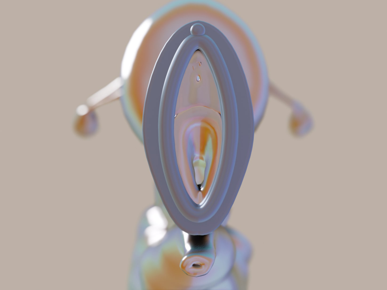
1. Pre-examination, the urogynaecologist will present an artefact that represents the vulva to the patient. One common issue with prolapse is that patients don’t have the vocabulary, or are embarrassed to talk about prolapse, even with a doctor.

2. Patients can use the artefact to gesture and explain where their prolapse is, and how far it protrudes outside of their body.

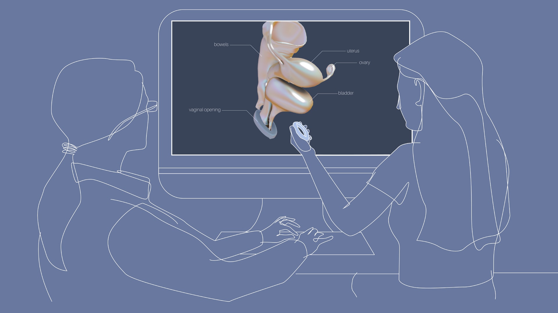
3. The urogynaecologist can refer to the AR software to explain internal anatomy, and how what a patient sees, is related to what’s going on inside of their body. 

Physical Artefact: Design Process, media item 1

I modelled an artefact in Fusion 360, printed it, and used it to create a silicone model in the RCA’s Future Material Lab. The first prototype of this iteration has been made out of Norcutt 40 Shore Silicone. The second will have a pattern on the back which is inlayed with a different color silicone to create a pattern that is recognisable to a Unity application. The top left image shows the vulva artefact created in Fusion 360, the centre image shows the model printed, ready to be used to create a mould, and the final image shows the result of the silicone mould. 

Augmented Reality: Design Process, media item 1

While the intention is for the physical artefacts to be distributed to urogynaecologists, there is also the design constraint of using as little physical material as possible, while creating as much impact as possible. As such, I have opted to only model what patients see with their own eyes, and keep anything they don’t ‘see’ as a virtual object. This use of AR also helps to maintain an abstraction between the inside and outside of the body.

I first used a sagittal cross section reference given to me by a urogynaecologist. Using Blender, I then modelled the three most important location markers: The bladder, uterus, and bowels. In a co-design workshop, I showed my first attempt to Dr. Pradhan, and then took notes on his critique. I have also consulted with tutors and patients about colour choice. For my initial prototype, I imported my model into Adobe Aero, and created an image tracking tag to superimpose the organ model over the physical vulva model. Unfortunately the image tracking capabilities of Aero are quite rudimentary, and it would only pick up letters and numbers. When trying to use a printed visual of the vulva, the software could not pick up the image. This is why I have chosen to use object tracking in Unity as the next step of the project. 

Thanks to Madrid-based 3D Artist, Juan Caso, I was able to fully realise my 3D prototypes with support structures such as ligaments and pelvic bones.

Illustrations, media item 1

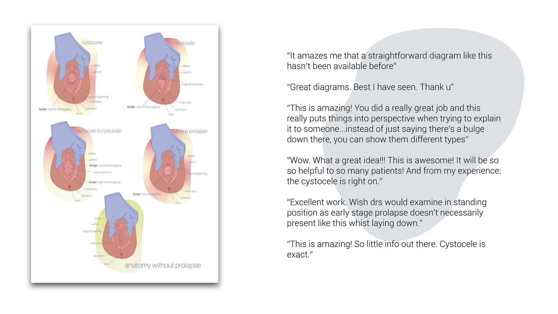
By researching the currently available prolapse diagrams, it’s clear that all illustrations are based on surgical illustrations. This imagery presents an inaccurate representation of prolapse to patients, who experience and see their bodies in a very different way to clinicians. I have been able to send proplapse visuals to a group of prolapse patients as well as clincians, and have been gathering qualitative feedback on the diagrams created. With this feedback, I will be able to redraw the current diagrams to be even more inclusive and understandable to patients. In the following weeks, Dr. Pradhan will also be showing the diagrams to his own patients to get feedback. The images developed were shared with the Let’s Talk about Prolapse Facebook group, which currently has over 15,000 members. The downloadable diagrams have been approved by over 200 women with prolapse.

Royal Commission of 1851